Masson染色法是利用两种或三种阴离子染料混合一起或先后作用对组织进行染色的一种方法,也是胶原纤维权威而经典的技术方法。染色时根据组织不同的渗透性能,选择分子大小不同的阴离子染料进行染色,便可把不同组织成分显色出来,主要适用于胶原蛋白和平滑肌的鉴别分析。
该产品具有无毒、环保,操作简捷,性能稳定,显色对比清晰,所染切片保存时间长且不易褪色等特点,广泛用于结缔组织、肌肉组织和胶原蛋白的研究等。
产品组分
| 组分编号 | 组分名称 | 产品编号/规格 | ||
| 60532ES58/80 mL | 60532ES66/160mL | 60532ES74/400mL | ||
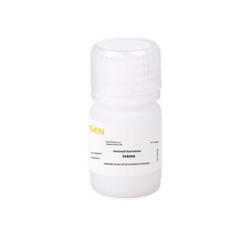
| 60532-A | Hematoxylin Stain Solution苏木素核染液 | 10 mL | 20 mL | 50 mL |
| 60532-B | Acid Fuchsin Stain Solution酸性品红浆染液 | 10 mL | 20 mL | 50 mL |
| 60532-C | Phosphomolybdic-phosphotungstic Acid Differentiation Solution磷钼酸分色液 | 10 mL | 20 mL | 50 mL |
| 60532-D | Aniline Blue Counterstain苯胺蓝复染液 | 10 mL | 20 mL | 50 mL |
| 60532-E | Washing Buffer 清洗液 | 10 mL×4 | 20 mL×4 | 50 mL×4 |
注意事项
1)为了您的安全和健康,请穿实验服并戴一次性手套操作。2)本产品仅作科研用途!
使用方法
1)染色前先用蒸馏水润湿玻片30-60 s。
2)加入适量苏木素核染液染色60 s左右,弃去,清洗液冲洗30 s左右。
3)加入适量酸性品红浆染液染色30-60 s左右,弃去,清洗液冲洗30 s左右。
4)加入适量磷钼酸分色液分色6-8 min,弃去。
5)加入适量苯胺蓝复染液染色5 min左右,弃去,用无水乙醇冲洗干净。
6)吹干后封片镜检(Yeasen Cat#36313ES60中性树胶封片剂)。
结果参考
胶原纤维、粘液、软骨呈蓝色;胞浆、肌肉、纤维素、神经胶质呈红色;胞核呈蓝紫色。
室温运输。室温保存,有效期12个月,开封后效期9个月。
Q:石蜡切片染色后还需要脱水吗?
A:苯胺蓝复染后用无水乙醇进行冲洗,之后直接吹干,封片即可。不用额外的再进行梯度脱水,透明等步骤。
Q:染色后发现染色结果不满意,如何处理?
A:可以把染色后的切片放到自来水中过夜(12h),等褪色后再进行重新染色。
Q:染色过程中发现片子不容易着色?
A:片子在染色前先用蒸馏水浸泡30-60s,可以促进染色。
Q:冲洗液用完了,配方是什么呢?
A:冲洗液配方是保密的,可以试下蒸馏水冲洗。
[1] Ji W, Hou B, Lin W, et al. 3D Bioprinting a human iPSC-derived MSC-loaded scaffold for repair of the uterine endometrium. Acta Biomater. 2020;116:268-284. doi:10.1016/j.actbio.2020.09.012(IF:7.242)
[2] Li B, Zhang Q, Sun J, Lai D. Human amniotic epithelial cells improve fertility in an intrauterine adhesion mouse model. Stem Cell Res Ther. 2019;10(1):257. Published 2019 Aug 14. doi:10.1186/s13287-019-1368-9(IF:6.832)
[3] Zheng Y, Li L, Bi X, Xue R. circPTP4A2-miR-330-5p-PDK2 Signaling Facilitates In Vivo Survival of HuMSCs on SF-SIS Scaffolds and Improves the Repair of Damaged Endometrium. Oxid Med Cell Longev. 2022;2022:2818433. Published 2022 May 6. doi:10.1155/2022/2818433(IF:6.543)
[4] Wang Y, Liu B, Wu P, et al. Dietary Selenium Alleviated Mouse Liver Oxidative Stress and NAFLD Induced by Obesity by Regulating the KEAP1/NRF2 Pathway. Antioxidants (Basel). 2022;11(2):349. Published 2022 Feb 10. doi:10.3390/antiox11020349(IF:6.313)
[5] Yang W, Zhang S, Ou T, et al. Interleukin-11 regulates the fate of adipose-derived mesenchymal stem cells via STAT3 signalling pathways. Cell Prolif. 2020;53(5):e12771. doi:10.1111/cpr.12771(IF:5.753)
[6] Wu D, Wang W, Li X, Yin B, Ma Y. Single-cell sequencing reveals the antifibrotic effects of YAP/TAZ in systemic sclerosis. Int J Biochem Cell Biol. 2022;149:106257. doi:10.1016/j.biocel.2022.106257(IF:5.652)
[7] Sun J, Pan Y, Li X, et al. Quercetin Attenuates Osteoporosis in Orchiectomy Mice by Regulating Glucose and Lipid Metabolism via the GPRC6A/AMPK/mTOR Signaling Pathway. Front Endocrinol (Lausanne). 2022;13:849544. Published 2022 Apr 25. doi:10.3389/fendo.2022.849544(IF:5.555)
[8] Shen H, Wang J, Min J, et al. Activation of TGF-β1/α-SMA/Col I Profibrotic Pathway in Fibroblasts by Galectin-3 Contributes to Atrial Fibrosis in Experimental Models and Patients. Cell Physiol Biochem. 2018;47(2):851-863. doi:10.1159/000490077(IF:5.500)
[9] Kim HS, Chen J, Wu LP, et al. Prevention of excessive scar formation using nanofibrous meshes made of biodegradable elastomer poly(3-hydroxybutyrate-co-3-hydroxyvalerate). J Tissue Eng. 2020;11:2041731420949332. Published 2020 Aug 23. doi:10.1177/2041731420949332(IF:5.352)
[10] Gu J, Chen C, Wang J, et al. Withaferin A Exerts Preventive Effect on Liver Fibrosis through Oxidative Stress Inhibition in a Sirtuin 3-Dependent Manner. Oxid Med Cell Longev. 2020;2020:2452848. Published 2020 Sep 24. doi:10.1155/2020/2452848(IF:5.076)
[11] Yang W, Zhang S, Zhu J, et al. Gut microbe-derived metabolite trimethylamine N-oxide accelerates fibroblast-myofibroblast differentiation and induces cardiac fibrosis. J Mol Cell Cardiol. 2019;134:119-130. doi:10.1016/j.yjmcc.2019.07.004(IF:5.055)
[12] Huang L, Xiang M, Ye P, Zhou W, Chen M. Beta-catenin promotes macrophage-mediated acute inflammatory response after myocardial infarction. Immunol Cell Biol. 2018;96(1):100-113. doi:10.1111/imcb.1019(IF:4.557)
[13] Xiang C, Zhu Y, Xu M, Zhang D. Fasudil Ameliorates Osteoporosis Following Myocardial Infarction by Regulating Cardiac Calcitonin Secretion [published online ahead of print, 2022 May 12]. J Cardiovasc Transl Res. 2022;10.1007/s12265-022-10271-8. doi:10.1007/s12265-022-10271-8(IF:4.132)
[14] Fan L, Liu Z, Zhang Z, et al. Identifying the clinical presentations, progression, and sequela of pelvic inflammatory disease through physiological, histological and ultrastructural evaluation of a rat animal model. Ann Transl Med. 2021;9(23):1710. doi:10.21037/atm-21-3345(IF:3.932)
[15] Zhou Y, Zhao Y, Du H, et al. Downregulation of CFTR Is Involved in the Formation of Hypertrophic Scars. Biomed Res Int. 2020;2020:9526289. Published 2020 Jan 2. doi:10.1155/2020/9526289(IF:2.276)
[16] Su Y, Tao X, Xu J. Protective effect of Alpinetin on rats with chronic obstructive pulmonary disease. Food Sci Nutr. 2020;8(12):6603-6611. Published 2020 Oct 25. doi:10.1002/fsn3.1952(IF:1.797)

