萤火虫萤光素酶(Firefly luciferase)是一种分子量约为61 kDa的蛋白,在ATP、镁离子和氧气存在的条件下,能够催化萤光素(luciferin)氧化成oxyluciferin,在luciferin氧化的过程中会发出波长为560 nm左右的生物荧光,该萤光可通过化学发光仪进行测定。检测原理如图所示:

图1:萤火虫萤光素酶检测原理图
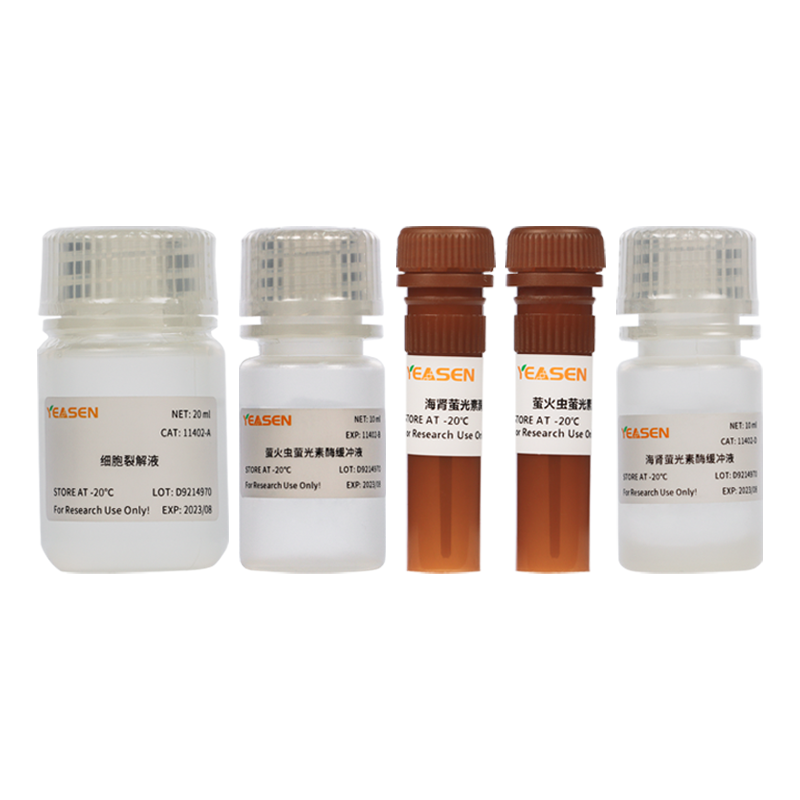
Luciferase Reporter Gene Assay kit是一闪光型萤火虫萤光素酶报告基因检测试剂盒,具有灵敏度高的特点,可以高灵敏的检测萤光素酶在哺乳动物细胞中的表达。
裂解能力更强:能够彻底裂解绝大部分种类细胞。
信号更强:能够精准检测弱启动子的表达。
灵敏度更高:可检测低至数百个细胞样本。
线性范围更广:线性检测范围超过酶浓度的8个数量级。
干冰运输。-20℃保存,有效期1年。
试剂盒内萤火虫萤光素酶检测试剂不能反复冻融,建议分装-20℃或-80℃保存。
Q:该试剂盒中裂解后一定要离心吗?
A:可以选做。
Q:测定时间为 10 s,测定间隔为 2 s,为什么这样推荐?如果这样设置,96 孔板这样设置,会导致每孔时间相差很大。仪器可以同时出数据。工程师建议设置动力学检测, 设置总时间。
A:不同仪器,设置时间可以不一样。单管手动检测,就按说明书设置会更好。
Q:酶标仪检测吗?
A:带有化学发光模块的可以。
Q:加入检测试剂后,产生的荧光稳定吗?多长时间内检测数据是准确的?
A:加入后海肾会有荧光猝灭的情况,越快越好,尽量 5min 内检测完成。
Q: 为啥11401试剂盒里要裂解细胞,40901这个单独的底物不需要啊?
A: 1、荧光素酶是表达在细胞内,是一种不存在于人类物种的水解酶(所以本底很低),它不能透膜。荧光素酶的底物是环状小分子,是疏水的,因而可以透膜的,因此只要存在荧光素酶表达,那必然是外源的,那么将底物加到细胞培养基中或打到体内均能得到信号。
2、报告基因体外检测需要细胞裂解是为了避免细胞转染异质性、底物渗透不均匀和信号不均一而强行匀质化的做法。体外报告基因检测是一个定量且灵敏度很高的实验,报告基因检测需要ATP和氧气的存在,裂解细胞是需要把表达的荧光素酶完全释放出来,能够有更好的反应,因为细胞内的氧气含量有限,如果不裂解,会导致信号一定程度的降低(有的裂解液也做了一些优化,比如额外加了ATP以提高信号等)。因此报告基因体外检测裂解细胞能够有更好的信号;
3、活体成像,检测仪器功率大,能够透过皮肤,细胞,有更强的穿透力,因此可以不用裂解细胞就可以检测到很强的荧光信号,还有就是活体成像检测比较糙,精准度都是相对的,
[1] Hao Y, Fan X, Shi Y, et al. Next-generation unnatural monosaccharides reveal that ESRRB O-GlcNAcylation regulates pluripotency of mouse embryonic stem cells. Nat Commun. 2019;10(1):4065. Published 2019 Sep 6. doi:10.1038/s41467-019-11942-y(IF:11.878)
[2] Wang X, Qin X, Yan M, et al. Loss of exosomal miR-3188 in cancer-associated fibroblasts contributes to HNC progression. J Exp Clin Cancer Res. 2019;38(1):151. Published 2019 Apr 8. doi:10.1186/s13046-019-1144-9(IF:11.161)
[3] Yin T, Liu Y, Yang M, Wang L, Zhou J, Huo M. Novel Chitosan Derivatives with Reversible Cationization and Hydrophobicization for Tumor Cytoplasm-Specific Burst Co-delivery of siRNA and Chemotherapeutics. ACS Appl Mater Interfaces. 2020;12(13):14770-14783. doi:10.1021/acsami.9b19373(IF:8.758)
[4] He Y, Qu J, Wei L, et al. Generation and Effect Testing of a SARS-CoV-2 RBD-Targeted Polyclonal Therapeutic Antibody Based on a 2-D Airway Organoid Screening System. Front Immunol. 2021;12:689065. Published 2021 Oct 18. doi:10.3389/fimmu.2021.689065(IF:7.561)
[5] Sun H, Chen D, Zhan S, et al. Design and Discovery of Natural Cyclopeptide Skeleton Based Programmed Death Ligand 1 Inhibitor as Immune Modulator for Cancer Therapy. J Med Chem. 2020;63(19):11286-11301. doi:10.1021/acs.jmedchem.0c01262(IF:6.205)
[6] Li S, Zhang L, Zhang G, et al. A nonautophagic role of ATG5 in regulating cell growth by targeting c-Myc for proteasome-mediated degradation. iScience. 2021;24(11):103296. Published 2021 Oct 16. doi:10.1016/j.isci.2021.103296(IF:5.458)
[7] Yang N, Xiong Y, Wang Y, et al. ADAP Y571 Phosphorylation Is Required to Prime STAT3 for Activation in TLR4-Stimulated Macrophages. J Immunol. 2021;206(4):814-826. doi:10.4049/jimmunol.2000569(IF:5.422)
[8] He S, Ma R, Liu Z, et al. Overexpression of BnaAGL11, a MADS-Box Transcription Factor, Regulates Leaf Morphogenesis and Senescence in Brassica napus. J Agric Food Chem. 2022;70(11):3420-3434. doi:10.1021/acs.jafc.1c07622(IF:5.279)
[9] Zhang RY, Zhou SH, He CB, et al. Adjuvant-Protein Conjugate Vaccine with Built-In TLR7 Agonist on S1 Induces Potent Immunity against SARS-CoV-2 and Variants of Concern. ACS Infect Dis. 2022;8(7):1367-1375. doi:10.1021/acsinfecdis.2c00259(IF:5.084)
[10] Li Y, Chen T, Wang W, et al. A high-efficiency Agrobacterium-mediated transient expression system in the leaves of Artemisia annua L. Plant Methods. 2021;17(1):106. Published 2021 Oct 16. doi:10.1186/s13007-021-00807-5(IF:4.993)
[11] Li D, Jiang K, Teng D, et al. Discovery of New Estrogen-Related Receptor α Agonists via a Combination Strategy Based on Shape Screening and Ensemble Docking. J Chem Inf Model. 2022;62(3):486-497. doi:10.1021/acs.jcim.1c00662(IF:4.956)
[12] Liu J, Chen X, Liu Y, et al. Characterization of SARS-CoV-2 worldwide transmission based on evolutionary dynamics and specific viral mutations in the spike protein. Infect Dis Poverty. 2021;10(1):112. Published 2021 Aug 21. doi:10.1186/s40249-021-00895-4(IF:4.388)
[13] Zhou FL, Li SC, Zhu Y, et al. Integrating yeast chemical genomics and mammalian cell pathway analysis [published correction appears in Acta Pharmacol Sin. 2020 May;41(5):729]. Acta Pharmacol Sin. 2019;40(9):1245-1255. doi:10.1038/s41401-019-0231-y(IF:4.010)
[14] Zhu G, Li X, Li J, et al. Arsenic trioxide (ATO) induced degradation of Cyclin D1 sensitized PD-1/PD-L1 checkpoint inhibitor in oral and esophageal squamous cell carcinoma. J Cancer. 2020;11(22):6516-6529. Published 2020 Sep 21. doi:10.7150/jca.47111(IF:3.565)
[15] Xie Y, Li X, Ge J. Cyclophilin A-FoxO1 signaling pathway in endothelial cell apoptosis. Cell Signal. 2019;61:57-65. doi:10.1016/j.cellsig.2019.04.014(IF:3.388)
[16] Wang Y, Bibi M, Min P, Deng W, Zhang Y, Du J. SOX2 promotes hypoxia-induced breast cancer cell migration by inducing NEDD9 expression and subsequent activation of Rac1/HIF-1α signaling. Cell Mol Biol Lett. 2019;24:55. Published 2019 Aug 22. doi:10.1186/s11658-019-0180-y(IF:3.367)
[17] Jian Y, Qiao Q, Tang J, Qin X. Origin recognition complex 1 regulates phospholipase Cδ1 to inhibit cell proliferation, migration and epithelial-mesenchymal transition in lung adenocarcinoma. Oncol Lett. 2022;24(2):252. Published 2022 Jun 10. doi:10.3892/ol.2022.13372(IF:2.967)
 相关产品
相关产品