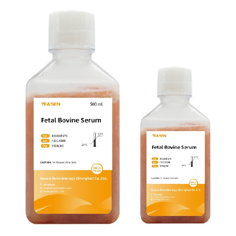
Yeasen胎牛血清(特级),经3次100 nm过滤和支原体、病毒筛查,经过严格检验测试后全自动化罐装,规范化生产,品质保证,含有丰富的细胞生长所需的营养成分,适用于培养大部分常规细胞系。
| 中文别名(Chinese Synonym) | 胎牛血清(特级) |
| 英文别名(English Synonym) | Fetal Bovine Serum Gold |
| 内毒素水平 | ≤5 EU/mL |
| 血红蛋白含量 | ≤0.02%(w/v) |
| 支原体检测 | 阴性 |
| 灭菌处理 | 三次100 nm过滤 |
产品选购指南
1. 适用于绝大多数常规细胞的培养,如常规肿瘤细胞、规模化生产细胞(293T、CHO、VERO等)、免疫细胞(RAW264.7、THP-1等)。其他娇贵细胞或难培养的细胞(如干细胞(如NSC、PB-MSCs等)、难培养的原代细胞(上皮细胞、内皮细胞、心肌细胞等)、其他难培养的细胞(如部分乳腺癌细胞、部分结直肠癌细胞、部分肺腺癌细胞、部分胶质瘤细胞等)建议选用40131ES、40132ES。
2. 多种生长因子,细胞培养状态稳定。
3. 纯天然制品,不含任何人为添加成分。
4. 为避免反复冻融,可以选购小规格(50 mL)产品,即用即取,方便快捷。10个50 mL产品可等同于500 mL产品。
产品优势
1. 兼容范围广——适用于多种常规细胞培养
2. 产品纯天然——对标进口胎牛血清,营养丰富,不含任何人为添加成分
3. 细胞状态好——多种生长因子,批次稳定,细胞培养状态稳定
4. 小规格便利——50 mL便携装,即用即取,避免反复冻融,避免沉淀产生,避免分装污染,方便快捷,产品升级不加价
5. 产品质控严——经过三次0.1 μM无菌过滤,有效去除微生物,通过支原体和病毒筛查检测
6. 内毒素极低——内毒素≤5 EU/mL,内毒素含量低,完全符合国家质量标准
7. 客户群体广——使用课题组数多达4000+,如中科院上海生命科学研究院,清华大学,浙江大学,上海交通大学等各大高校和研究院
8. 发表文献多——客户发表的文章引用影响因子累计高达1000+
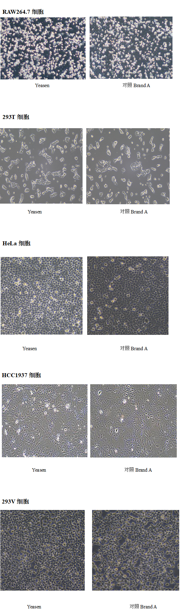
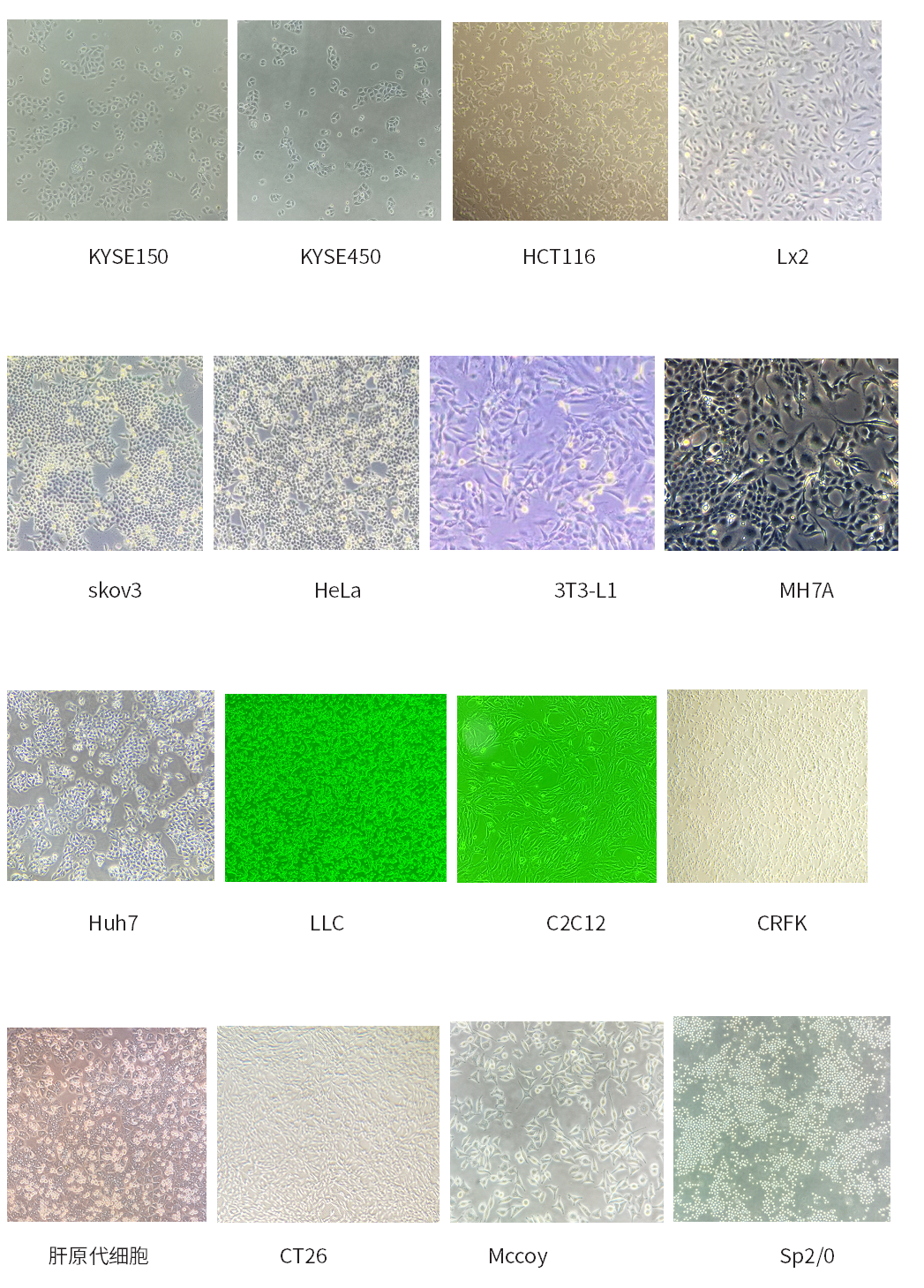
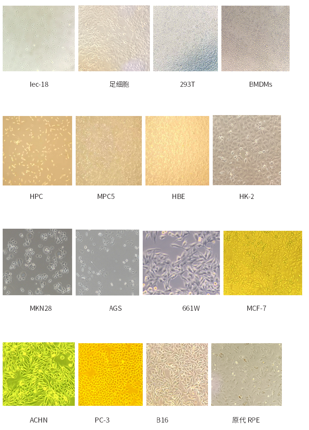
产品应用案例(细胞培养)
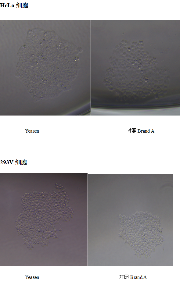
产品应用案例(单克隆实验)
客户使用本产品发表的科研文献(部分)
[1] Hu S, Peng L, Xu C, Wang Z, Song A, Chen FX. SPT5 stabilizes RNA polymerase II, orchestrates transcription cycles, and maintains the enhancer landscape. Mol Cell. 2021;81(21):4425-4439.e6. doi:10.1016/j.molcel.2021.08.029 (IF:17.970)
[2] Teng KX, Niu LY, Xie N, Yang QZ. Supramolecular photodynamic agents for simultaneous oxidation of NADH and generation of superoxide radical. Nat Commun. 2022;13(1):6179. Published 2022 Oct 19. doi:10.1038/s41467-022-33924-3 (IF:17.694)
[3] Tu J, Li W, Yang S, et al. Single-Cell Transcriptome Profiling Reveals Multicellular Ecosystem of Nucleus Pulposus during Degeneration Progression. Adv Sci (Weinh). 2022;9(3):e2103631. doi:10.1002/advs.202103631 (IF:16.806)
[4] Zhang W, Liu J, Li X, et al. Precise Chemodynamic Therapy of Cancer by Trifunctional Bacterium-Based Nanozymes. ACS Nano. 2021;15(12):19321-19333. doi:10.1021/acsnano.1c05605 (IF:15.881)
[5] Zhao C, Xie Y, Xu L, et al. Structures of a mammalian TRPM8 in closed state. Nat Commun. 2022;13(1):3113. Published 2022 Jun 3. doi:10.1038/s41467-022-30919-y (IF:14.919)
[6] Cai Z, Zhang Y, Zhang W, et al. Arsenic retention in erythrocytes and excessive erythrophagocytosis is related to low selenium status by impaired redox homeostasis. Redox Biol. 2022;52:102321. doi:10.1016/j.redox.2022.102321 (IF:11.799)
[7] Zhai Y, Liu M, Yang T, et al. Self-activated arsenic manganite nanohybrids for visible and synergistic thermo/immuno-arsenotherapy. J Control Release. 2022;350:761-776. doi:10.1016/j.jconrel.2022.08.054 (IF:11.467)
[8] Liu M, Chen C, Yu J, et al. The gelatin-based liquid marbles for cell cryopreservation. Mater Today Bio. 2022;17:100477. Published 2022 Oct 31. doi:10.1016/j.mtbio.2022.100477 (IF:10.761)
[9] Li S, Zhang J, Qian S, et al. S100A8 promotes epithelial-mesenchymal transition and metastasis under TGF-β/USF2 axis in colorectal cancer. Cancer Commun (Lond). 2021;41(2):154-170. doi:10.1002/cac2.12130 (IF:10.392)
产品应用
| 胎牛血清(特级)40130ES适用细胞系(不完全统计) | |||
| 序号 | 物种 | 细胞名称 | 中文名称 |
| 1 | 人 | CCRF-CEM | 人急性淋巴细胞白血病T淋巴细胞 |
| 2 | 人 | A-431 | 人皮肤鳞癌细胞 |
| 3 | 小鼠 | SP2/0 | 小鼠骨髓瘤细胞 |
| 4 | 人 | CFPAC-1 | 人胰腺癌细胞 |
| 5 | 人 | 6T-CEM | 人T细胞白血病细胞 |
| 6 | 人 | T84 | 人结直肠癌细胞 |
| 7 | 人 | Hela 229 | 人宫颈癌细胞 |
| 8 | 人 | Huh-7 | 人肝癌细胞 |
| 9 | 人 | PC-3 | 人前列腺癌细胞 |
| 10 | 人 | 2BS | 人肺二倍体细胞 |
| 11 | 人 | Jurkat E6-1 | 人急性T淋巴细胞白血病细胞 |
| 12 | 人 | HCCLM3 | 高转移人肝癌细胞 |
| 13 | 人 | MRC-5 | 人肺二倍体细胞 |
| 14 | 人 | MOLM-13 | 人急性髓系白血病细胞 |
| 15 | 人 | NCI-H209 | 人小细胞肺癌细胞 |
| 16 | 人 | MSC | 人脐带间充质干细胞 |
| 17 | 人 | NCI-H1650 | 人非小细胞肺癌细胞 |
| 18 | 人 | NCI-H1395 | 人肺腺癌细胞 |
| 19 | 大鼠 | RH-35 | 大鼠肝癌细胞 |
| 20 | 人 | BT-474 | 人乳腺导管癌细胞 |
| 21 | 人 | PANC-1 | 人胰腺癌细胞 |
| 22 | 人 | SH-SY5Y | 人神经母细胞瘤 |
| 23 | 人 | HCC 94 | 人子宫鳞癌细胞 |
| 24 | 人 | ME-180 | 人子宫颈表皮癌细胞 |
| 25 | 人 | 786-O | 人肾细胞腺癌细胞 |
| 26 | 人 | MG-63 | 人骨肉瘤细胞 |
| 27 | 人 | RBE | 人肝胆管癌细胞 |
| 28 | 小鼠 | MKWFCs | 小鼠表皮角质形成细胞 |
| 29 | 人 | BT-549 | 人乳腺管癌细胞 |
| 30 | 人 | COLO 320DM | 人结直肠腺癌细胞 |
| 31 | 人 | ES-2 | 人卵巢透明细胞癌 |
| 32 | 人 | HAL-01 | 人淋巴细胞白血病细胞 |
| 33 | 人 | HCT-8 | 人回盲肠癌细胞 |
| 34 | 人 | Hep3B2-1-7 | 人肝癌细胞 |
| 35 | 人 | Hs-683 | 人脑神经胶质瘤细胞 |
| 36 | 人 | K-562 | 人慢性髓原白血病细胞 |
| 37 | 人 | WI-38 | 人胚肺成纤维细胞 |
| 38 | 大鼠 | NRK | 大鼠肾细胞 |
| 39 | 大鼠 | PC-12(低分化) | 大鼠肾上腺嗜铬细胞瘤细胞(低分化) |
| 40 | 大鼠 | PC-12(高分化) | 大鼠肾上腺嗜铬细胞瘤细胞(高分化) |
| 41 | 人 | KG-1 | 人急性骨髓性白血病细胞 |
| 42 | 人 | ARPE-19 | 人视网膜上皮细胞 |
| 43 | 人 | Farage | 人B淋巴瘤细胞 |
| 44 | 人 | NK92 | 人恶性非霍奇金淋巴瘤患者的自然杀伤细胞 |
| 45 | 人 | SW-13 | 人肾上腺皮质小细胞癌细胞 |
| 46 | 人 | A3 | 人T淋巴细胞白血病细胞 |
| 47 | 人 | ZR-75-30 | 人乳腺癌细胞 |
| 48 | 人 | SNU-387 | 人肝癌细胞 |
| 49 | 人 | SK-MEL-5 | 人皮肤黑色素瘤细胞 |
| 50 | 人 | MCF7 | 人乳腺癌细胞 |
| 51 | 人 | MDA-MB-436 | 人乳腺腺癌细胞 |
| 52 | 人 | MKN74 | 人胃癌细胞 |
| 53 | 人 | NCI-H1437 | 人肺癌细胞 |
| 54 | 人 | NCI-H524 | 人非小细胞肺癌细胞 |
| 55 | 人 | NCI-H23 | 人肺癌细胞 |
| 56 | 人 | NCI-H661 | 人大细胞肺癌细 |
| 57 | 人 | LM3 | 肝癌细胞株 |
| 58 | 人 | NCI-H508 | 人结肠癌细胞 |
| 59 | 人 | PA-1 | 人卵巢畸胎瘤细胞 |
| 60 | 人 | RG2 [D74] | 大鼠胶质瘤细胞 |
| 61 | 小鼠 | bEnd.3 | 小鼠脑微血管内皮细胞 |
| 62 | 人 | MHCC-97H | 人高转移潜能肝癌细胞 |
| 63 | 小鼠 | MB49 | 小鼠膀胱癌细胞 |
| 64 | 小鼠 | ID8 | 小鼠卵巢上皮癌细胞 |
| 65 | 小鼠 | HC11 | 小鼠乳腺上皮细胞 |
| 66 | 小鼠 | MC38 | 小鼠结肠癌细胞 |
| 67 | 小鼠 | AML12 | 小鼠肝细胞 |
| 68 | 人 | hccc-9810 | 人胆管细胞型肝癌细胞 |
| 69 | 人 | Hep G2 | 人肝癌细胞 |
| 70 | 人 | Calu-6 | 人退行性癌细胞 |
| 71 | 人 | MM.1S | 人多发性骨髓瘤细胞 |
| 72 | 小鼠 | P19 | 小鼠畸胎瘤细胞 |
| 73 | 人 | PC-9 | 人肺癌细胞 |
| 74 | 人 | NCI-H226 | 人肺鳞癌细胞 |
| 75 | 人 | NCI-H929 | 人骨髓瘤细胞 |
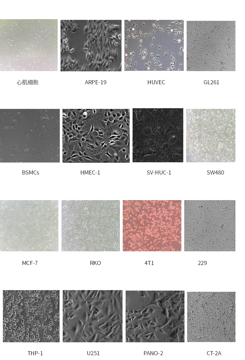
客户使用本产品的数据展示

运输方式:干冰运输;
保存方式:-20℃至-10℃可保存5年。
【注】一旦解冻,血清应该保存在2℃到8℃冰箱,存储时间不宜超过6周。如果需要长期保存,建议将血清在无菌环境下先进行分装,后重新冷冻保存,避免反复冻融。
解冻的方式(二选一)
1) 将血清从-20℃存储条件下取出,放置于2-8℃冰箱中过夜,使其部分溶解,然后在室温条件下使其全部融解,溶解过程中须不时的摇动瓶身混合里面的液体。建议采取此种方式解冻更优。
2) 直接将血清从-20℃取出后,放置于37℃水浴中,不断摇晃瓶身使其加快解冻和混合。解冻以后不要在37℃水浴放置太长时间。
【注】如果不晃动瓶身,当温度超过40℃的时候沉积在瓶底的物质有可能会发生蛋白变形,出现沉淀。如果出现沉淀,建议可将血清分装到无菌离心管中,400~600 g(如500 g)离心5 min,取血清上清液,加入到基础培养基中,再一起过滤,全程保证无菌环境即可。
注意事项
1)使用过程中,避免反复冻融,避免紫外线照射。
2)避免血清在37℃环境中时间过长。
3)避免血清在2~8℃条件下长期保存,可以将血清无菌分装后,置于-20℃的环境中长期保存。
4)避免血清在室温条件下过长时间放置。
5)为了您的安全和健康,请穿实验服并戴一次性手套操作。
6)本产品仅作科研用途!
Q: 请问我们的血清拿到之后还需要热灭活吗?
A: 现在商业化的血清产品,基本上出厂之前都经过了灭活处理,所以一般情况下,就不需要再操作一次灭活了。
Q: 解冻后血清中有悬浮物质/絮状沉淀,因怎样处理?
A: 血清中沉淀物的出现有许多种原因,但最普遍的原因是由于血清中脂蛋白的变性所造成,而血纤维蛋白(形成凝血的蛋白之一)在血清解冻后,也会存在于血清中,亦是造成沉淀物的主要原因之一。但这些絮状沉淀物,并不影响血清本身的质量。
去除这些絮状沉淀物,可以将血清分装至无菌离心管内,以400g稍微离心10-15mins去除沉淀。我们不建议以过滤的方法去除这些絮状物,因为它可能会阻塞您过滤膜。建议在使用血清的时候,注意正确的血清解冻步骤,并尽量避免灭活血清及长时间的将血清置于高温环境中。
Q:为什么我们的血清看着比较黄,竞品公司血清比较红?
A: 血红蛋白的原因,血红蛋白含量比较高,血清就会比较红,血红蛋白含量较少,所以会比较黄,血清本质是黄的。
[1] Hu S, Peng L, Xu C, Wang Z, Song A, Chen FX. SPT5 stabilizes RNA polymerase II, orchestrates transcription cycles, and maintains the enhancer landscape. Mol Cell. 2021;81(21):4425-4439.e6. doi:10.1016/j.molcel.2021.08.029(IF:17.970)
[2] Tu J, Li W, Yang S, et al. Single-Cell Transcriptome Profiling Reveals Multicellular Ecosystem of Nucleus Pulposus during Degeneration Progression. Adv Sci (Weinh). 2022;9(3):e2103631. doi:10.1002/advs.202103631(IF:16.806)
[3] Zhang W, Liu J, Li X, et al. Precise Chemodynamic Therapy of Cancer by Trifunctional Bacterium-Based Nanozymes. ACS Nano. 2021;15(12):19321-19333. doi:10.1021/acsnano.1c05605(IF:15.881)
[4] Zhao C, Xie Y, Xu L, et al. Structures of a mammalian TRPM8 in closed state. Nat Commun. 2022;13(1):3113. Published 2022 Jun 3. doi:10.1038/s41467-022-30919-y(IF:14.919)
[5] Cai Z, Zhang Y, Zhang W, et al. Arsenic retention in erythrocytes and excessive erythrophagocytosis is related to low selenium status by impaired redox homeostasis. Redox Biol. 2022;52:102321. doi:10.1016/j.redox.2022.102321(IF:11.799)
[6] Li S, Zhang J, Qian S, et al. S100A8 promotes epithelial-mesenchymal transition and metastasis under TGF-β/USF2 axis in colorectal cancer. Cancer Commun (Lond). 2021;41(2):154-170. doi:10.1002/cac2.12130(IF:10.392)
[7] Yang X, Qiu Q, Liu G, et al. Traceless antibiotic-crosslinked micelles for rapid clearance of intracellular bacteria. J Control Release. 2022;341:329-340. doi:10.1016/j.jconrel.2021.11.037(IF:9.776)
[8] Wang X, Qi Y, Wang Z, et al. RPAP2 regulates a transcription initiation checkpoint by inhibiting assembly of pre-initiation complex. Cell Rep. 2022;39(4):110732. doi:10.1016/j.celrep.2022.110732(IF:9.423)
[9] Ma Z, Zhang Y, Zhang J, et al. Ultrasmall Peptide-Coated Platinum Nanoparticles for Precise NIR-II Photothermal Therapy by Mitochondrial Targeting. ACS Appl Mater Interfaces. 2020;12(35):39434-39443. doi:10.1021/acsami.0c11469(IF:8.758)
[10] Sun Q, Ye Y, Gui A, et al. MORTALIN-Ca2+ axis drives innate rituximab resistance in diffuse large B-cell lymphoma. Cancer Lett. 2022;537:215678. doi:10.1016/j.canlet.2022.215678(IF:8.679)
[11] Wang Y, Sun Q, Ye Y, et al. FGF-2 signaling in nasopharyngeal carcinoma modulates pericyte-macrophage crosstalk and metastasis. JCI Insight. 2022;7(10):e157874. Published 2022 May 23. doi:10.1172/jci.insight.157874(IF:8.315)
[12] Wang Y, Sun Q, Ye Y, et al. FGF-2 signaling in nasopharyngeal carcinoma modulates pericyte-macrophage crosstalk and metastasis. JCI Insight. 2022;7(10):e157874. Published 2022 May 23. doi:10.1172/jci.insight.157874(IF:8.315)
[13] Wang Y, Sun Q, Ye Y, et al. FGF-2 signaling in nasopharyngeal carcinoma modulates pericyte-macrophage crosstalk and metastasis. JCI Insight. 2022;7(10):e157874. Published 2022 May 23. doi:10.1172/jci.insight.157874(IF:8.315)
[14] Liu Z, Tao C, Li S, et al. circFL-seq reveals full-length circular RNAs with rolling circular reverse transcription and nanopore sequencing. Elife. 2021;10:e69457. Published 2021 Oct 14. doi:10.7554/eLife.69457(IF:8.146)
[15] Cao J, Peng X, Li H, et al. Ultrasound-assisted continuous-flow synthesis of PEGylated MIL-101(Cr) nanoparticles for hematopoietic radioprotection. Mater Sci Eng C Mater Biol Appl. 2021;129:112369. doi:10.1016/j.msec.2021.112369(IF:7.328)
[16] Ma H, Lin J, Li L, et al. Formaldehyde reinforces pro-inflammatory responses of macrophages through induction of glycolysis. Chemosphere. 2021;282:131149. doi:10.1016/j.chemosphere.2021.131149(IF:7.086)
[17] Chen Y, Chen Y, Jiang X, et al. Vascular Adventitial Fibroblasts-Derived FGF10 Promotes Vascular Smooth Muscle Cells Proliferation and Migration in vitro and the Neointima Formation in vivo. J Inflamm Res. 2021;14:2207-2223. Published 2021 May 25. doi:10.2147/JIR.S305204(IF:6.922)
[18] Wang Y, Zhao M, Li W, et al. BMSC-Derived Small Extracellular Vesicles Induce Cartilage Reconstruction of Temporomandibular Joint Osteoarthritis via Autotaxin-YAP Signaling Axis. Front Cell Dev Biol. 2021;9:656153. Published 2021 Apr 1. doi:10.3389/fcell.2021.656153(IF:6.684)
[19] Liu W, Wu Z, Yu Y, et al. Functional Evaluation of KEL as an Oncogenic Gene in the Progression of Acute Erythroleukemia. Oxid Med Cell Longev. 2022;2022:5885342. Published 2022 Jan 30. doi:10.1155/2022/5885342(IF:6.543)
[20] Wang G, Wang H, Jin Y, et al. Galactooligosaccharides as a protective agent for intestinal barrier and its regulatory functions for intestinal microbiota. Food Res Int. 2022;155:111003. doi:10.1016/j.foodres.2022.111003(IF:6.475)
[21] Zhi W, Li S, Wan Y, Wu F, Hong L. Short-term starvation synergistically enhances cytotoxicity of Niraparib via Akt/mTOR signaling pathway in ovarian cancer therapy [published correction appears in Cancer Cell Int. 2022 Mar 21;22(1):131]. Cancer Cell Int. 2022;22(1):18. Published 2022 Jan 11. doi:10.1186/s12935-022-02447-8(IF:5.722)
[22] Wang Q, Zhu Y, Li Z, et al. Up-regulation of SPC25 promotes breast cancer. Aging (Albany NY). 2019;11(15):5689-5704. doi:10.18632/aging.102153(IF:5.515)
[23] Liu XF, Zhu XD, Feng LH, et al. Physical activity improves outcomes of combined lenvatinib plus anti-PD-1 therapy in unresectable hepatocellular carcinoma: a retrospective study and mouse model. Exp Hematol Oncol. 2022;11(1):20. Published 2022 Apr 4. doi:10.1186/s40164-022-00275-0(IF:5.133)
[24] Gao Z, Wang T, Li R, et al. The discovery of a novel series of potential ERRα inverse agonists based on p-nitrobenzenesulfonamide template for triple-negative breast cancer in vivo. J Enzyme Inhib Med Chem. 2022;37(1):125-134. doi:10.1080/14756366.2021.1995728(IF:5.051)
[25] Liu ZZ, Duan XX, Yuan MC, et al. Glucagon-like peptide-1 receptor activation by liraglutide promotes breast cancer through NOX4/ROS/VEGF pathway. Life Sci. 2022;294:120370. doi:10.1016/j.lfs.2022.120370(IF:5.037)
[26] Wu Z, Wang Q, Yang H, et al. Discovery of Natural Products Targeting NQO1 via an Approach Combining Network-Based Inference and Identification of Privileged Substructures. J Chem Inf Model. 2021;61(5):2486-2498. doi:10.1021/acs.jcim.1c00260(IF:4.956)
[27] Yang X, Miao BS, Wei CY, et al. Lymphoid-specific helicase promotes the growth and invasion of hepatocellular carcinoma by transcriptional regulation of centromere protein F expression. Cancer Sci. 2019;110(7):2133-2144. doi:10.1111/cas.14037(IF:4.751)
[28] Dai Y, Li Y, Lin G, et al. Non-pathogenic grass carp reovirus infection leads to both apoptosis and autophagy in a grass carp cell line [published online ahead of print, 2022 Jun 21]. Fish Shellfish Immunol. 2022;127:681-689. doi:10.1016/j.fsi.2022.06.022(IF:4.581)
[29] Qin F, Zhang W, Zhang M, et al. Adipose-Derived Stem Cells Improve the Aging Skin of Nude Mice by Promoting Angiogenesis and Reducing Local Tissue Water. Aesthet Surg J. 2021;41(7):NP905-NP913. doi:10.1093/asj/sjab001(IF:4.283)
[30] Luo H, Zheng J, Chen Y, et al. Utility Evaluation of Porcine Enteroids as PDCoV Infection Model in vitro. Front Microbiol. 2020;11:821. Published 2020 Apr 23. doi:10.3389/fmicb.2020.00821(IF:4.236)
[31] Ma H, Ding Z, Xie Y, et al. Methylglyoxal produced by tumor cells through formaldehyde-enhanced Warburg effect potentiated polarization of tumor-associated macrophages. Toxicol Appl Pharmacol. 2022;438:115910. doi:10.1016/j.taap.2022.115910(IF:4.219)
[32] Peng X, Wang K, Zhang C, et al. The mitochondrial antioxidant SS-31 attenuated lipopolysaccharide-induced apoptosis and pyroptosis of nucleus pulposus cells via scavenging mitochondrial ROS and maintaining the stability of mitochondrial dynamics. Free Radic Res. 2021;55(11-12):1080-1093. doi:10.1080/10715762.2021.2018426(IF:4.148)
[33] Niu Y, Liu F, Wang X, et al. miR-183-5p Promotes HCC Migration/Invasion via Increasing Aerobic Glycolysis. Onco Targets Ther. 2021;14:3649-3658. Published 2021 Jun 4. doi:10.2147/OTT.S304117(IF:4.147)
[34] Sun J, Zhou YQ, Xu BY, et al. STING/NF-κB/IL-6-Mediated Inflammation in Microglia Contributes to Spared Nerve Injury (SNI)-Induced Pain Initiation [published online ahead of print, 2021 Nov 2]. J Neuroimmune Pharmacol. 2021;10.1007/s11481-021-10031-6. doi:10.1007/s11481-021-10031-6(IF:4.147)
[35] Duan H, Lei Z, Xu F, et al. PARK2 Suppresses Proliferation and Tumorigenicity in Non-small Cell Lung Cancer. Front Oncol. 2019;9:790. Published 2019 Aug 23. doi:10.3389/fonc.2019.00790(IF:4.137)
[36] Liu Q, Tian R, Yu P, Shu M. miR-221/222 suppression induced by activation of the cAMP/PKA/CREB1 pathway is required for cAMP-induced bidirectional differentiation of glioma cells. FEBS Lett. 2021;595(22):2829-2843. doi:10.1002/1873-3468.14208(IF:4.124)
[37] Zeng WJ, Lu C, Shi Y, et al. Initiation of stress granule assembly by rapid clustering of IGF2BP proteins upon osmotic shock. Biochim Biophys Acta Mol Cell Res. 2020;1867(10):118795. doi:10.1016/j.bbamcr.2020.118795(IF:4.105)
[38] Pan H, Chai W, Liu X, Yu T, Sun L, Yan M. DYNC1H1 regulates NSCLC cell growth and metastasis by IFN-γ-JAK-STAT signaling and is associated with an aberrant immune response. Exp Cell Res. 2021;409(1):112897. doi:10.1016/j.yexcr.2021.112897(IF:3.905)
[39] Qin F, Huang J, Zhang W, et al. The Paracrine Effect of Adipose-Derived Stem Cells Orchestrates Competition between Different Damaged Dermal Fibroblasts to Repair UVB-Induced Skin Aging. Stem Cells Int. 2020;2020:8878370. Published 2020 Dec 17. doi:10.1155/2020/8878370(IF:3.869)
[40] Wang L, Hu D, Xie B, Xie L. Blockade of Myd88 signaling by a novel MyD88 inhibitor prevents colitis-associated colorectal cancer development by impairing myeloid-derived suppressor cells. Invest New Drugs. 2022;40(3):506-518. doi:10.1007/s10637-022-01218-6(IF:3.850)
[41] Xie H, Zhang C, Zhang J, et al. An in vitro cell model to study microglia activation in diabetic retinopathy. Cell Biol Int. 2022;46(1):129-138. doi:10.1002/cbin.11710(IF:3.612)
[42] Li X, Wang X, Miao L, Guo Y, Yuan R, Tian H. Design, synthesis, and neuroprotective effects of novel hybrid compounds containing edaravone analogue and 3-n-butylphthalide ring-opened derivatives. Biochem Biophys Res Commun. 2021;556:99-105. doi:10.1016/j.bbrc.2021.03.171(IF:3.575)
[43] Zhu Y, Wu H, Yang X, Xiong Z, Zhao T, Gan X. LINC00514 facilitates cell proliferation, migration, invasion, and epithelial-mesenchymal transition in non-small cell lung cancer by acting on the Wnt/β-catenin signaling pathway. Bioengineered. 2022;13(5):13654-13666. doi:10.1080/21655979.2022.2084246(IF:3.269)
[44] Wang XH, Gao JW, Bao JP, et al. GATA4 promotes the senescence of nucleus pulposus cells via NF-κB pathway. Arch Gerontol Geriatr. 2022;101:104676. doi:10.1016/j.archger.2022.104676(IF:3.250)
[45] Huang D, Xiong M, Xu X, et al. Bile acids elevated by high-fat feeding induce endoplasmic reticulum stress in intestinal stem cells and contribute to mucosal barrier damage. Biochem Biophys Res Commun. 2020;529(2):289-295. doi:10.1016/j.bbrc.2020.05.226(IF:2.985)
[46] Ma H, Song X, Huang P, et al. Myricetin protects natural killer cells from arsenite induced DNA damage by attenuating oxidative stress and retaining poly(ADP-Ribose) polymerase 1 activity. Mutat Res Genet Toxicol Environ Mutagen. 2021;865:503337. doi:10.1016/j.mrgentox.2021.503337(IF:2.873)
[47] Wang D, Xue Z, Wu H, Shi G, Feng S, Zhao L. Hepatoprotective effect and structural analysis of Hedysarum polysaccharides in vivo and in vitro [published online ahead of print, 2022 Apr 28]. J Food Biochem. 2022;e14188. doi:10.1111/jfbc.14188(IF:2.720)
[48] Xiao L, Zhang S, Zheng Q, Zhang S. Dysregulation of KIF14 regulates the cell cycle and predicts poor prognosis in cervical cancer: a study based on integrated approaches. Braz J Med Biol Res. 2021;54(11):e11363. Published 2021 Sep 3. doi:10.1590/1414-431X2021e11363(IF:2.590)
[49] Li T, Liu Y, Zhang Q, Sun W, Dong Y. A steroid-induced osteonecrosis model established using an organ-on-a-chip platform. Exp Ther Med. 2021;22(4):1070. doi:10.3892/etm.2021.10504(IF:2.447)
[50] Zheng GD, Hu PJ, Chao YX, et al. Nobiletin induces growth inhibition and apoptosis in human nasopharyngeal carcinoma C666-1 cells through regulating PARP-2/SIRT1/AMPK signaling pathway. Food Sci Nutr. 2019;7(3):1104-1112. Published 2019 Feb 10. doi:10.1002/fsn3.953(IF:1.747)
[51] Ying J, Huang HH, Zhang MM, Chen JF. Up-regulation of SOCS4 promotes cell proliferation and migration in esophageal squamous cell carcinoma. Transl Cancer Res. 2021;10(5):2416-2427. doi:10.21037/tcr-21-700(IF:1.241)